- About
-
Research
- Agronomy and farming systems
-
Agricultural crop research
-
Research projects - agriculture
- About SASSA-SAI
- BioBoost
- Biomass Connect
- CTP for Sustainable Agricultural Innovation
- Climate Ready Beans - workshop presentations (March 2022)
- Crop diversity HPC cluster
- Designing Future Wheat
- Final project workshop
- Get involved
- List of materials
- News and updates
- Partners
- Rustwatch
- The Sentinel Crop Disease Surveillance Network
- The research team
- UK Cereal Pathogen Virulence Survey
- UK wheat varieties pedigree
- Weed management - IWM Praise
- Crop breeding
- Crop characterisation
- Data sciences
- Genetics and pre-breeding
- Plant biotechnology
- Plant pathology and entomology
- Resources
-
Research projects - agriculture
-
Horticultural crop research
-
Research projects - horticulture
- Augmented Berry Vision
- BEESPOKE
- Boosting brassica nutrition in smart growing systems
- CTP for Fruit Crop Research
- Develop user-friendly nutrient demand models
- Egg laying deterrents for spotted wing drosophila
- Enhancing the nutritional quality of tomatoes
- Improving berry harvest forecasts and productivity
- Improving vineyard soil health through groundcover management
- Intelligent growing systems
- Knowledge transfer for sustainable water use
- POME: Precision Orchard Management for Environment
- RASCAL
- STOP-SPOT
- UV-Robot
- Crop science and production systems
- Genetics, genomics and breeding
- Pest and pathogen ecology
- Field vegetables and salad crops
- Plum Demonstration Centre
- The WET Centre
- Viticulture and Oenology
-
Research projects - horticulture
- Crop Science Centre
-
Services
- Analytical Services
- Business Development
- Commercial trial services
- Digital
- Membership
- Plant breeding
- Plant characterisation
- Seed certification
-
Training
-
Technical agronomy training
- Advanced crop management of bulb onions
- Advanced crop management of vegetable brassicas
- Advanced nutrient management for combinable crops
- Benefits of cover crops in arable systems
- Best practice agronomy for cereals and oilseed rape
- Developing a Successful Strategy for Spring Crops
- Disease Management and Control in Cereal Crops
- Exploring regenerative agriculture
- Improving Soil Organic Matter and Farm Carbon Management
- Incorporating SFI options into your rotation
- Protected Environment Horticulture – Best Practice
- Techniques for better pest management in combinable crops
- Exploring regenerative agriculture
- Quantitative methods in plant breeding
- Seed certification & crop inspectors
-
Technical agronomy training
- News & Views
-
Event Hub
- Book your place
-
Variety choice
- POSTER: Grass and forage variety choice (2024)
- POSTER: Oilseed rape variety choice (2024)
- POSTER: Pulse variety choice (2024)
- POSTER: Wheat variety blends (2024)
-
Variety choice - archive
- Video: NIAB at Cereals 2020
- Video: Group 1 winter wheat variety choice (2020)
- Video: Group 2 winter wheat variety choice (2020)
- Video: Group 3 winter wheat variety choice (2020)
- Video: Group 4 winter wheat variety choice (2020)
- Video: NIAB @ the Cereals Event (2020)
- Video: Winter wheat varieties and Rustwatch (2020)
- POSTER: Oilseed rape variety choice (2023)
- POSTER: Pulse variety choice (2023)
- Crop nutrition and management
-
Disease management
- POSTER: Disease control in winter wheat (2024)
- POSTER: UKCPVS disease monitoring (2024)
- POSTER: Finding new sources of Septoria resistance (2024)
- POSTER: Fungicide resistance research (2024)
- POSTER: Detecting air-borne pathogens (2024)
- POSTER: Disease control in barley (2024)
- POSTER: Oilseed rape diseases (2024)
- Disease management - 2023
-
Disease management - 2022
- POSTER: Monitoring yield response to fungicides (2022)
- Poster: Pathogen diagnostics (2022)
- POSTER: fungicide management (2022)
- POSTER: Septoria surveillance (2022)
- POSTER: Monitoring disease race changes in yellow rust (2022)
- POSTER: Improving chocolate spot resistance (2022)
- POSTER: Developing an early warning system for wheat rust (2022)
- Disease management - 2021
- Disease management - 2020
- Centre for High Carbon Capture Cropping
- Oilseeds, pulses & break crops
-
Soils and farming systems
- POSTER: Checking soil health - across space and time (2024)
- POSTER: Checking soil health - step by step (2024)
- POSTER: Checking soil health - step by step (2024)
- POSTER: Soil structure and organic matter (2024)
- POSTER: Novel wheat genotypes for regen-ag (2024)
- POSTER: Assessing the soil at the Cereals Event (2024)
- POSTER: Impact of prolonged rainfall on soil structure (2024)
-
Soils and farming systems 2020-23
- POSTER: Saxmundham experimental site (2023)
- POSTERS: Changing soil management practices (2022)
- Poster: Monitoring natural enemies & pollinators (2021)
- STAR Online Seminar 2021
- Video: New Farming Systems project (2021)
- Video: Saxmundham Experimental Site (2021)
- Video: Soil health assessment (2021)
- Poster: Controlled Traffic Farming (2020)
- VIDEO: Conservation agriculture (2020)
- VIDEO: Great Soils; soil sampling guidelines (2020)
- Video: Conservation agriculture - a farmer's view (2020)
- Video: Cover crops, living mulch and leys (2020)
- Video: Improving soil (2020)
- Webinar: CFE soils, water, wildlife & profit (2020)
- Webinar: Sensors & biostimulants (2020)
- Webinar: Soil health & circular economy (2020)
- Poster: Cover crops (2020)
- POSTER: Soil & agronomic monitoring study (2024)
- POSTER: The impact of rotations & cultivations (2024)
- Poster: Soil invertebrates within arable rotations (2024)
- Climate change & sustainability
-
Weed management
- Poster: Weed seed predation in regen-ag (2024)
- POSTER: Understanding the hierarchy of black-grass control (2023)
- POSTER: herbicide resistance surveys (2023)
- POSTER: Mode of action (2023)
- POSTER: Inter-row cultivation for black-grass control (2022)
- POSTER: Emerging weed threats (2022)
- Poster: Management of Italian ryegrass (2021)
- Video: Inter-row cultivations for black-grass control (2020)
- Poster: Grass weed management (2020)
- Video: Wild oat seed sampling (2020)
-
Crop genetics
- POSTER: Diversity enriched wheat (2024)
- POSTER: Genetics of wheat flag leaf size (2024)
- POSTER: Wheat yield stability (2024)
- POSTER: Domestic chickpea production (2024)
- Poster: Traits for future cereal crops (2022)
- POSTER: wild wheat fragment lines (2022)
- POSTER: Improving phenotyping in crop research (2022)
- PRESENTATION: Plant breeding for regen ag
- Video: Tour NIAB's 2020 wheat breeding plots
- Poster: Designing Future Wheat (2020)
- Fruit crops
- Potatoes
-
Seed certification and variety testing
- POSTER: Wheat DUS (2024)
- POSTER: Innovation in variety testing (2024)
- POSTER: AI and molecular markers for soft fruit (2024)
- POSTER: Barley crop identification (2023)
- POSTER: Herbage grass crop identification (2023)
- POSTER: Herbage legume crop identification (2024)
- POSTER: Minor cereal crop inspecting (2023)
- POSTER: Pulse crop identification (2023)
- POSTER: Wheat crop identification (2023)
Symptoms and recognition (Apple canker)

Cankers
- These initially appear as sunken areas of bark around buds, leaf scars, shoot bases or open wounds.
- As the canker develops the centre dies and bark flakes off.
- Old lesions show as flaky dark brown strips of bark surrounded by swollen wound tissue.
- Red or white fruiting bodies may be present.
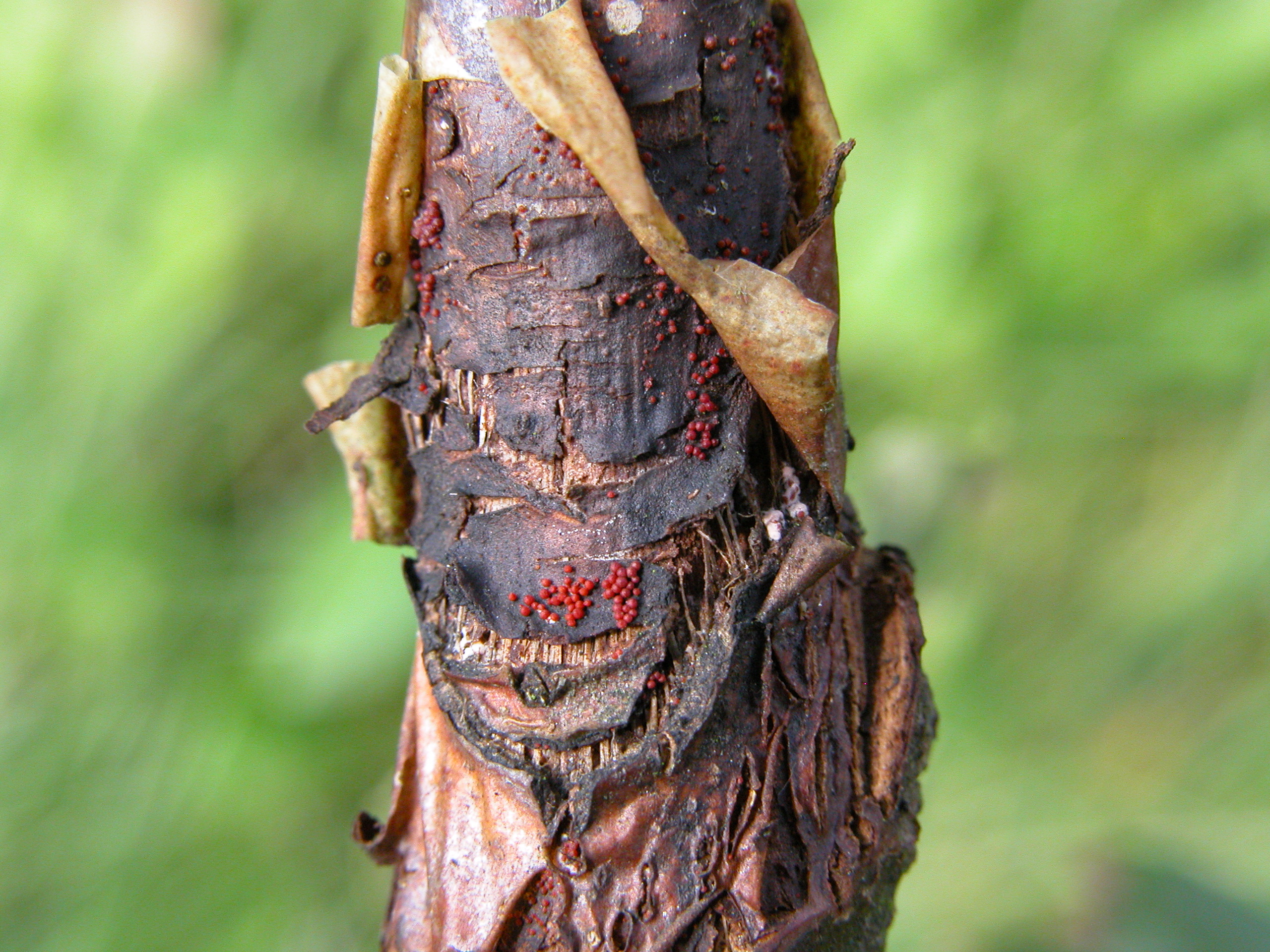
- Young cankers, particularly those on young shoots, tend to have white fruiting bodies (conidial spore masses – asexual state).
- White fruiting bodies tend to be present in the summer and early autumn, whereas red fruiting bodies or perithecia (sexual state) are present in autumn, winter and spring.
- Shoot dieback due to canker is common in canker prone orchards in early summer.
- Cankers on wood may result in wilting and/or browning of leaves and blossoms on the branch above the canker, which may occur even before the branch is girdled.
- Trees infected with canker show brown staining in the wood when cut which can usually be traced back to a canker.
- Both the leaf symptoms and wood staining are thought to be due to the production of toxin by the N. ditissima fungus.
- Similarly, blossoms wilting as a result of Neonectria canker located further down the branch can be confused with blossoms wilting or dying due to blossom wilt, fireblight or bud moth.
Diagnosis of cankers
| Disease/problem | Canker description | Fruiting bodies | Canker location |
|---|---|---|---|
| Blossom wilt (Monilinia laxa f.sp. mali) | Brown/cracked, distinct light/dark zones of infection | Grey pustules in spring | Fruiting spur, base of fruiting spur, branch |
| Brown rot (Monilinia fructigena) | Brown/cracked, distinct light/dark zones of infection | Buff pustules in summer | Fruiting spur, base of fruiting spur, branch |
| Apple canker (Neonectria ditissima) | Distinct cankered areas. Initially sunken areas around bud, leaf scar, shoot base, or wound. Older cankers, flaky brown bark strips surrounded by swollen tissue. Sometimes papery bark on cankered young shoots. | White/creamy pustules especially on young cankers in summer. Red pin-head sized fruiting bodies in autumn and winter, which can be confused with eggs of fruit tree red spider mite. | Young shoots causing shoot dieback, shoot bases, branches of all ages, tree crotches, branch angles, main trunk especially of young trees, rootstock. |
| Fireblight (Erwinia amylovora) | Cankers indistinct, associated with dieback. Cankered area slightly sunken and darker than healthy tissue with separating crack. Internal tissue water-soaked with red/brown streaks. | Milky bacterial ooze. | Shoot dieback. Disease progression to branch |
| Coral spot (Nectria cinnabarina) | Cankers indistinct, associated with twig or branch dieback | Pinkish pustules in summer. Pinhead –sized red fruiting bodies in winter | Shoot /branch dieback. Often associated with pruning snags. |
| Papery bark (physiological) | Initially pale blister-like swellings which eventually develop into peeling papery bark. | On young shoots and older branches. Often associated with excessive soil moisture. | |
| Silver leaf (Chondrostereum purpureum) | Associated with pruning wound. Blistering and papery bark near wound. Affected wood if cut is discoloured. Foliage on tree or tree part is silvered. | Small bracket fruiting bodies (creamy-coloured above and purple below) arise on affected tree parts once they die. | On large branches, associated with pruning wounds especially major tree restructuring. |
| Perennial (Neofabraea – formerly Gloeosporium) canker (Pezicula malicorticis) | Distinct cankers. Initially small circular brownish / purplish spots that develop into elliptical cankers separated from healthy tissue by crack. Bark in affected area sloughs off | Cream-coloured fruiting bodies develop on the cankers. | Associated with wounds, either pruning, frost cracks etc. |
Diagnosis of wilting dying blossoms
| Disease/Problem | Blossom symptom | Fruiting bodies | Smell | Other symptoms |
|---|---|---|---|---|
| Blossom wilt (Monilinia laxa f.sp. mali) | Wilting/brown,Internal browning/necrosis | Grey pustules on infected parts | Fetid smell, similar to scent of sweet chestnut flowers | Disease progression into spur and branch forming cankers |
| Apple canker (Neonectria ditissima) | Wilting/brown, no internal browning | None | None | Nectria canker somewhere on branch with wilting blossoms |
| Fireblight (Erwinia amylovora) | Wilting/brown, internal browning/necrosis | Milky bacterial ooze on infected flower parts | None | Disease progression into spur and branch, possible further ooze |
| Bud moth (Spilonota ocellana) | Wilting/brown blossom. Hollow | None | None | Evidence of internal mining, caterpillar and frass |
Fruit rot
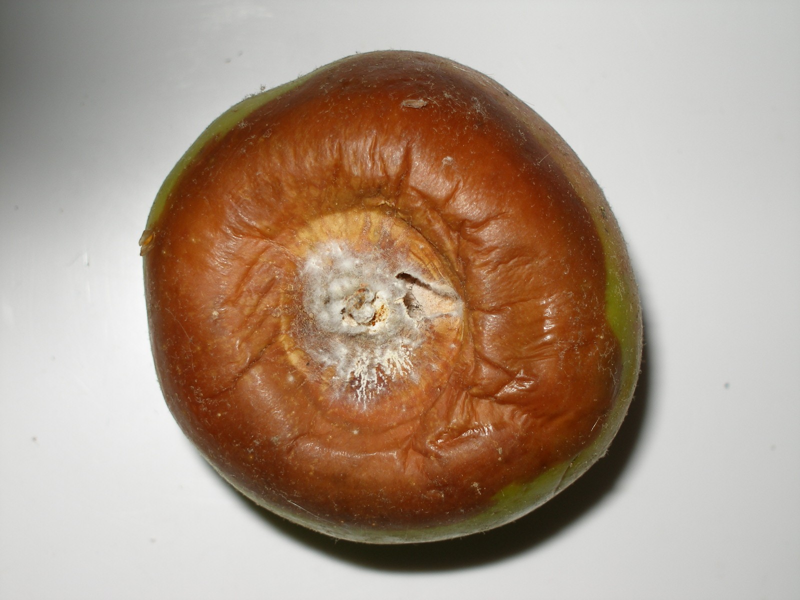
- The fruit rot occurs on the eye, the stalk end or on the cheek.
- The rots are soft, slightly sunken, with the rotted part easily scooped out from the sound flesh.
- Eye rots are visible in the orchard from early summer as well as in store. They are usually brown in colour with white/creamy cobwebby sporulating pustules visible on mature rots.
- Cheek and stalk-end rots only appear in store and are circular, brown with pale brown centres.
- Neonectria rots appear in cold-stored fruit from late December onwards and increase in incidence the longer the fruit is stored.
- The rot colour depends on variety and storage conditions.
- Rots on fruit stored in low oxygen tend to be green in colour with very little sporulation.
- Those in higher oxygen storage tend to be brown with white/creamy sporing pustules.
Other problems that may be confused with apple canker
Cankers
Many other fungi cause cankers on apple trees.
- The most common are blossom wilt, brown rot, perennial (Neofabraea – formerly Gloeosporium) canker.
- Neonectria cankers can usually be readily distinguished from these because they are distinct cankers, rather than die back, and because of the presence of white or red fruiting bodies.
Fruit rots
Neonectria fruit rot can be confused with rots caused by Neofabraea – formerly Gloeosporium spp. or Penicillium spp. These rots similarly occur at the stalk, cheek and calyx end of the fruit.
- Those caused by Penicillium spp. are usually squashier, paler green in colour with pure white or turquoise-green spore pustules present.
- Rots caused by Neofabraea – formerly Gloeosporium species may only be distinguishable by microscopic examination of spores, if present, or culturing the fungus on to agar media.
Contact
Niab
Park Farm
Villa Road
Histon
Cambridge
CB24 9NZ, UK
Tel: +44(0)1223 342200
Email: [email protected]


Niab Agronomy
Membership

© NIAB 2025