- About
-
Research
- Agronomy and farming systems
-
Agricultural crop research
-
Research projects - agriculture
- About SASSA-SAI
- CTP for Sustainable Agricultural Innovation
- Climate Ready Beans - workshop presentations (March 2022)
- Crop diversity HPC cluster
- Final project workshop
- Get involved
- List of materials
- News and updates
- Partners
- The research team
- UK Cereal Pathogen Virulence Survey
- UK wheat varieties pedigree
- Weed management - IWM Praise
- Crop breeding
- Crop characterisation
- Data sciences
- Genetics and pre-breeding
- Plant biotechnology
- Plant pathology and entomology
- Resources
-
Research projects - agriculture
- Horticultural crop research
- Crop Science Centre
- Research Projects
- Research Publications
-
Services
- Analytical Services
- Business Development
- Commercial trial services
- Membership
- Plant breeding
- Plant characterisation
- Seed certification
-
Training
-
Technical agronomy training
- Advanced crop management of bulb onions
- Advanced crop management of vegetable brassicas
- Advanced nutrient management for combinable crops
- Benefits of cover crops in arable systems
- Best practice agronomy for cereals and oilseed rape
- Developing a Successful Strategy for Spring Crops
- Disease Management and Control in Cereal Crops
- Incorporating SFI options into your rotation
- Protected Environment Horticulture – Best Practice
- Techniques for better pest management in combinable crops
- Crop inspector and seed certification
- Licensed seed sampling
-
Technical agronomy training
- News & Views
- Events
-
Knowledge Hub
- Alternative and break crops
-
Crop genetics
- POSTER: Diversity enriched wheat (2025)
- POSTER: Genetics of wheat flag leaf size (2024)
- POSTER: Wheat yield stability (2024)
- Poster: Traits for future cereal crops (2022)
- POSTER: wild wheat fragment lines (2022)
- POSTER: Improving phenotyping in crop research (2022)
- PRESENTATION: Plant breeding for regen ag
- Poster: Designing Future Wheat (2020)
- Crop nutrition
-
Crop protection
- POSTER: Understanding the hierarchy of black-grass control (2025)
- POSTER: Emerging weed threats (2025)
- POSTER: Disease control in barley (2025)
- Poster: Weed seed predation in regen-ag (2024)
- POSTER: Disease control in winter wheat (2025)
- POSTER: Mode of action (2023)
- POSTER: Inter-row cultivation for black-grass control (2022)
- POSTER: UKCPVS winter wheat yellow rust in spring 2025 (2025)
- Poster: Management of Italian ryegrass (2021)
- POSTER: UKCPVS winter wheat rusts - 2024/25 review (2025)
- POSTER: UKCPVS disease monitoring and the benefit to UK growers (2025)
- POSTER: Diagnosing and scoring crop disease using AI (2025)
- POSTER: Finding new sources of Septoria resistance (2024)
- POSTER: Fungicide resistance research (2024)
- POSTER: Detecting air-borne pathogens (2024)
- POSTER: Oilseed rape diseases (2024)
- POSTER: Fungicide resistance research (2024)
- POSTER: Improving chocolate spot resistance (2022)
- Poster: Pathogen diagnostics (2022)
- Fruit
- Regen-ag & sustainability
-
Seed certification
- POSTER: Wheat DUS (2024)
- POSTER: Innovation in variety testing (2024)
- POSTER: AI and molecular markers for soft fruit (2024)
- POSTER: Barley crop identification (2023)
- POSTER: Herbage grass crop identification (2023)
- POSTER: Herbage legume crop identification (2024)
- POSTER: Minor cereal crop inspecting (2023)
- POSTER: Pulse crop identification (2023)
- POSTER: Wheat crop identification (2023)
-
Soils and farming systems
- POSTER: Checking soil health - across space and time (2024)
- POSTER: Checking soil health - step by step (2024)
- POSTERS: Changing soil management practices (2022)
- Poster: Monitoring natural enemies & pollinators (2021)
- POSTER: Soil structure and organic matter (2024)
- POSTER: Novel wheat genotypes for regen-ag (2024)
- Video: New Farming Systems project (2021)
- Video: Saxmundham Experimental Site (2021)
- POSTER: Impact of prolonged rainfall on soil structure (2024)
- POSTER: Soil & agronomic monitoring study (2024)
- POSTER: The impact of rotations & cultivations (2024)
- VIDEO: Great Soils; soil sampling guidelines (2020)
- Poster: Soil invertebrates within arable rotations (2024)
- VIDEO: Soil health assessment (2021)
- POSTER: Saxmundham - modern P management learnings
- POSTER: Saxmundham - 125 years of phosphorus management
- Poster: Soil phosphorus - availability, uptake and management (2025)
- POSTER: Morley long term experiments (2025)
- POSTER: Exploiting novel wheat genotypes for regen-ag (2025)
- Video: Saxmundham Experimental Site (2021)
- Varieties
PRESS RELEASE: A microscopic view of ergot infecting wheat wins science photography award
Proving that agricultural science can be visually stunning as well as informative an image of the ergot fungus (Claviceps purpurea) growing inside wheat stigma hairs is a winning entry at the Wellcome Image Awards, an annual science and medical photography competition.
The image shows the ergot hyphae growing through the same tissues of the wheat flower as a growing pollen tube would do during pollination. The wheat stigma hairs are only 25 micrometres, or 0.025mm, wide.
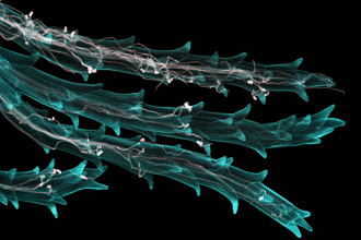
The photo was taken by Dr Anna Gordon, a molecular plant pathologist at NIAB, and Dr Fernan Federici from Cambridge University, with a confocal microscope 30 hours after the wheat plants had been infected. It is part of a BBSRC-funded project investigating the biological interaction at a transcriptomic level between ergot and wheat during infection and looking for sources of natural resistance.
Contact
Niab
Park Farm Campus
Villa Road
Histon
Cambridge
CB24 9AT, UK
Tel: +44(0)1223 342200
Email: [email protected]


Niab Agronomy
Membership